INTRODUCTION
1 ANATOMY AND PHYSIOLOGY OF THE DIGESTIVE SYSTEM
Section I. Anatomy of the
Digestive System
Section II. Functions and
Stages of the Digestive Process
Exercises
2 PHYSICAL ASSESSMENT OF THE DIGESTIVE SYSTEM
Exercises
3 DISEASES AND DISORDERS OF THE GASTROINTESTINAL
SYSTEM
Section I. Diseases/Disorders
of the Upper Gastrointestinal System
Section II. Hernias
Section III.
Diseases/Disorders of the Lower Gastrointestinal System
Section IV. Diseases/Disorders
of the Accessory Organs
Exercises
4 INGESTED POISONS
Exercises
5 NASOGASTRIC INTUBATION
Section I. Preliminary Steps
Section II. Procedure for
Inserting the Nasogastric Tube
Section III. Procedure for
Removing the Nasogastric Tube
Exercises
6 ABDOMINAL TRAUMA
Exercises
7 HEPATITIS
Exercises
------------------------------
LESSON 1
ANATOMY AND PHYSIOLOGY OF THE DIGESTIVE SYSTEM
Section I. ANATOMY OF THE DIGESTIVE SYSTEM
1-1. INTRODUCTION
a. Food is Essential to Life. Food is necessary
for the chemical reactions that take place in every body cell; for
example, formation of new enzymes, cell structures, bone, and all
other parts of the body that give the energy to supply the body's
needs.
Most of the foods we eat are just too large to pass
through the plasma membranes of the cells. The process of breaking
down food molecules for the body's cells to use is called digestion,
and the organs which work together to perform this function are termed
the digestive system.
b. Regulation of Food Intake. How much food we
eat is regulated by two sensations--hunger and appetite. When we crave
food in general, we are experiencing hunger, and when we want a
specific food, the correct term is appetite. The stronger of the two
sensations is hunger which is accompanied by a stronger feeling of
discomfort.
The hypothalamus is the control center for food
intake. There are a cluster of nerve cells in the lateral hypothalamus
(the appetite center) which send impulses causing a person to want to
eat. Another cluster of nerve cells tell the person he has had enough.
These cells are located in the medial hypothalamus and
called the satiety center. A person's food intake must be regulated to
prevent the digestive tract from becoming too full. The upper
digestive tract expands to let food enter the tract. Receptors in the
walls of the digestive tract are stimulated and send signals to the
satiety center, signals that tell the person he is full. He stops
taking in food, and the contents of the digestive tract are digested.
c. Digestive Processes. Five basic activities
help the digestive system prepare for use by the cells. These
activities are ingestion, peristalsis, digestion, absorption, and
defecation.
(1) Ingestion. Taking into the body of food, drink, or
medicines by mouth.
(2) Peristalsis. Alternating contraction and
relaxation of the walls of a tubular structure by which food is move
along the digestive tract.
(3) Digestion. The processes by which food is broken
down chemically and mechanically for the body's use. In chemical
digestion, catabolic reactions break down protein, lipid, and large
carbohydrate molecules we have eaten into smaller molecules which can
be used by the body's cells. Mechanical digestion refers to the
various movements which aid chemical digestion. Examples of such
movements are the chewing of food by teeth and the churning of food by
the smooth muscles of the stomach and the small intestine.
(4) Absorption. The taking up of digested food from the digestive
tract into the cardiovascular and lymphatic systems for distribution
to the body's cells.
(5) Defecation. The discharge of indigestible
substances from the body.
d. Organization of Digestive Organs. The
digestive organs are commonly divided into two main groups: the
gastrointestinal (GI) tract (also called the alimentary canal) and the
accessory structures.
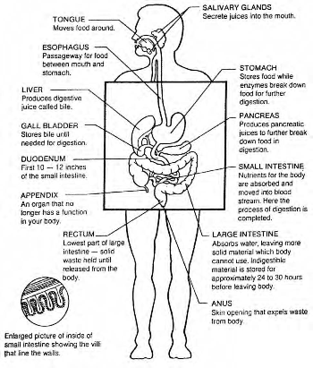
(1) The gastrointestinal (GI) tract. The
gastrointestinal tract is a continuous tube which extends from the
mouth to the anus and which runs through the ventral body cavity. The
tube is about 30 feet long in a cadaver and a little shorter in a
living person because the tube's wall muscles are toned. From the time
food is eaten until it is digested and eliminated, it is in the
gastrointestinal tract. Muscular contractions in the walls of the GI
tract churn the food breaking it into usable molecules. The organs
which make up the gastrointestinal tract are the mouth, pharynx,
esophagus, stomach, small intestine, and large intestine. These organs
are sometimes referred to as the primary organs of the digestive
system.
(2) Accessory structures. These structures include the
teeth, tongue, salivary glands, liver, gallbladder, and pancreas.
Except for the teeth and the tongue, all the structures lie outside
the continuous tube which is the gastrointestinal tract. Secretions
that aid in the chemical breakdown of food are produced and stored by
these structures. Eventually, such secretions are released into the GI
tract through ducts in the body.
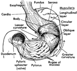
(3) General histology (structure of tissues). The
gastrointestinal wall has the same basic tissue arrangement from the
mouth to the anus. There are four coats (also called tunics): the
mucosa, submucosa, muscularis, and serosa (adventitia). The mucosa,
the inner tissue layer, contains blood and lymph vessels which carry
nutrients to other tissues and also protects the rest of the body
against disease. The submucosa is made up of loose connective tissue
and binds the mucosa to the next layer which is the muscularis.
Skeletal muscle in the muscularis of the mouth, pharynx, and esophagus
produce voluntary swallowing. The outer layer of tissue is the serosa.
NOTE: Remember that the GI tract carries food which often
contains bacteria.
From The
Gastrointestinal System


